Dr. Alvin Jeon recently presented a case of a 20 year old M who presented to the hospital with chest pain. Given the atypical nature of the chest pain, a CT chest was obtained and revealed a large anterior mediastinal mass that was ultimately found to be a thymoma.
This diagnosis sparked an excellent discussion regarding Anterior Mediastinal Masses.
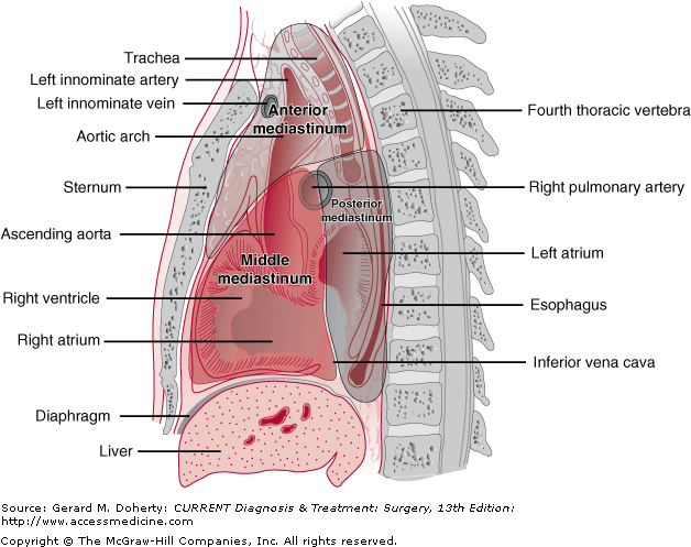
When approaching mediastinal masses, it’s best to compartmentalize into: Anterior, middle or Posterior mediastinum – each with their own unique differential!
For Anterior Mediastinal Masses, I always remember the the 5-Ts:
- Terrible Lymphadenopathy
- Thymic Tumors
- Teratoma
- Thyroid Mass
- Thoracic Aortic Aneurysm
Thymomas typically comprise about 20% of mediastinal masses. The presenting symptoms can vary but can range from asymptomatic to exhibiting localized symptoms (ex. Chest pain). Less commonly, thymomas can present with paraneoplastic syndromes:
- Myasthenia Gravis: about half the patients with thymomas will have symptoms consistent with myasthenia gravis (both men and women)
- Pure Red Cell Aplasia: less common (5-15%), but still important paraneoplastic syndrome. This is an autoimmune condition where there is hypo proliferation of the erythrocyte precursors in the bone marrow. More common in older women
Fortunately for our patient, he did not have any paraneoplastic syndromes and he is scheduled for a thymectomy!
References:
Berry, MF., Bogard, AJ., Approach to the adult patient with a mediastinal mass, In: UpToDate, Muller, NL., Midthun, DE., Vallières, E., UpToDate, Waltham, MA, 2020
Bird, S. Pathogenesis of myasthenia gravis, In: UpToDate, Shefner, JM. , UpToDate, Waltham, MA, 2020